CT scanning reveals downward tephra injectites

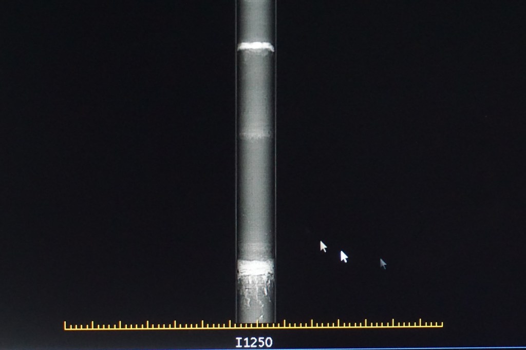
This week we analysed the internal structure of the sediment cores we collected at Rotoroa, Rotokaeo, and Waiwhakareke using a medical CT scanner at Hamilton Radiology. This method provides a first estimation about whether or not seismites (tephras deformed by earthquake activity) are present in the sedimentary record. Next week we plan to cut the cores lengthwise for detailed sediment description and sampling.

Sediment cores were scanned using a new medical CT scanner at Hamilton Radiology. From left to right: Dr Vicki Moon, Dr Max Kluger, Jordanka Chaneva, and Nic Ross
X-ray image of a sediment core, which shows downward injection structures from (tentatively) Rotorua tephra layer into underlying organic-rich host sediments
CT image of sediment core. Upper left: Downward injection structures from (tentatively) Tuhua and Mamaku tephras

Leave a Reply

Fill in your details below or click an icon to log in:

WordPress.com Logo

You are commenting using your WordPress.com account. Log Out /  Change )

Facebook photo

You are commenting using your Facebook account. Log Out /  Change )

Connecting to %s